
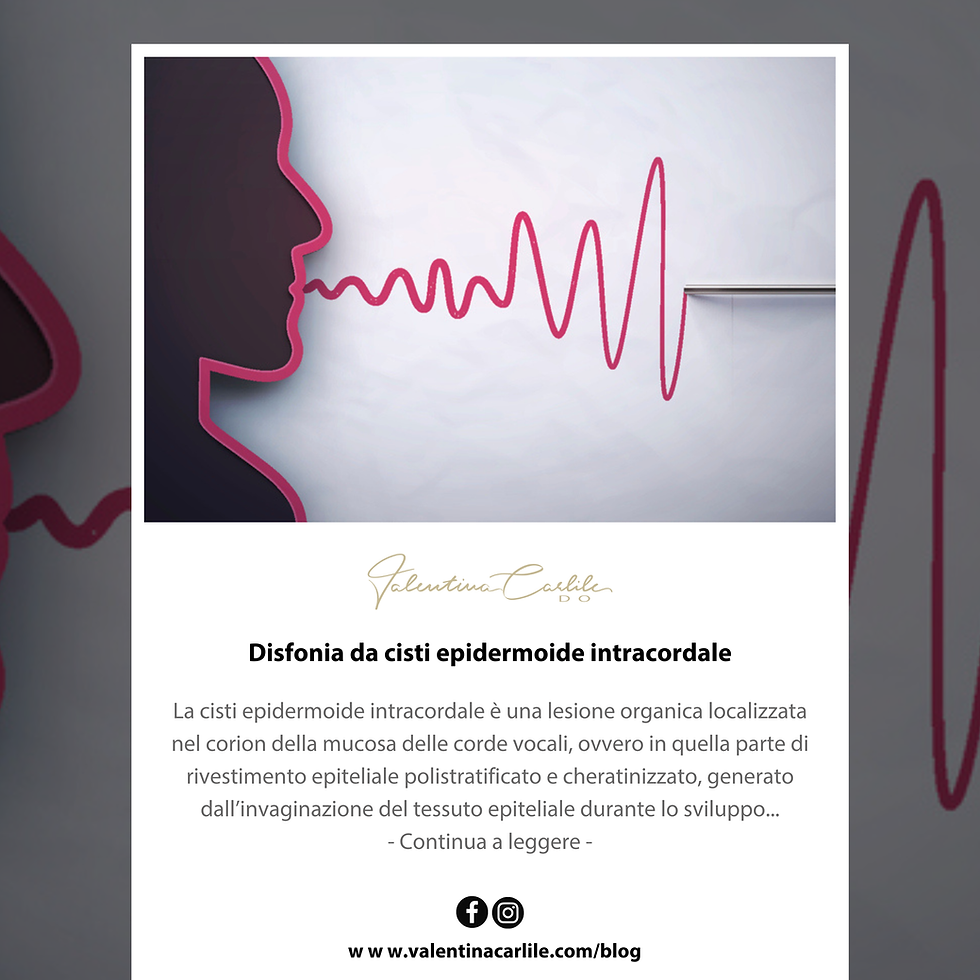
Intracordal epidermoid cyst-related dysphonia
- Valentina Carlile DO

- Nov 25, 2025
- 2 min read
The intracordal epidermoid cyst is an organic lesion located in the corium of the vocal cord mucosa, that is, in the part of the stratified keratinized epithelial lining, generated by the invagination of epithelial tissue during embryonic development. Occasionally, due to its constant growth and vocal trauma, it can evolve into an open cyst and create a groove in the affected vocal cord.
It is not always a round and whitish lesion, but tends to adapt to the layers of the cord, acquiring longitudinal shapes, which can make it difficult to identify.
Symptoms depend on the degree of mucosal wave involvement. If the cyst is small and superficial, it can go unnoticed for years, perhaps causing periodic dysphonia coinciding with the start of work or activities that require vocal overload. If the cyst is larger and adheres to the vocal ligament, it blocks the movement of the body mass, usually presenting from childhood as a hoarse voice, lacking resonance and modulation. These are children who produce voice with great effort and tend to vocal fatigue.
Laryngoscopy shows a rounded lesion beneath the mucosa. Edema and vascular dilation are almost always present in the cord, which usually converges toward the cyst. Sometimes the cyst is not visible, and only edema and vascular dilation are observed, known as vascular corditis. The presence of an isolated polyp (sentinel polyp) may also indicate an underlying cystic lesion.
Stroboscopy reveals an asymmetric, delayed wave with reduced amplitude and incomplete closure. At other times, the mucosa, instead of being homogeneous, progresses from the posterior to the anterior part. Associated signs such as monocorditis, mild edema, unilateral nodules of the cordal edge, and even asymmetric nodules may be observed.
Valentina Carlile - Osteopath specializing in Osteopathy for Voice and Speech Disorders since 2002. For information and bookings, visit the Contact page.


Osservo una sintesi equilibrata delle variabili analitiche in questo articolo. Le affermazioni rimangono supportate da prove. Il sito Web offre una spiegazione contestuale più approfondita del problema. L'ampiezza dell'analisi è ampliata dagli ecosistemi di servizi interattivi.